A) posterior gray column
B) dorsal gray ganglion
C) posterior white column
D) posterior gray horn
E) anterior gray horn
G) A) and C)
Correct Answer

verified
Correct Answer
verified
Multiple Choice
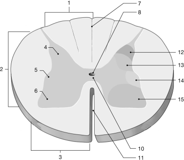
 Figure 13-1 The Spinal Cord
Use Figure 13-1 to answer the following questions:
-What is the function of the structure labeled "12"?
A) control of skeletal muscle
B) somatic sensory receiving
C) visceral sensory receiving
D) control of visceral effectors
E) ascending pathway
G) A) and D)
Correct Answer

verified
Correct Answer
verified
Short Answer
A(n)________ reflex has at least one interneuron placed between the sensory and motor neurons.
Correct Answer

verified
Correct Answer
verified
Multiple Choice
The ventral root of a spinal nerve contains
A) axons of motor neurons.
B) axons of sensory neurons.
C) cell bodies of motor neurons.
D) cell bodies of sensory neurons.
E) interneurons.
G) All of the above
Correct Answer

verified
Correct Answer
verified
Essay
Sometimes,when it is difficult to initiate a knee-jerk reflex by tapping the patellar tendon,a patient will be asked to voluntarily make a fist.Then the reflex will be easily evoked.What does this illustrate about the relation between voluntary and involuntary reflex movement?
Correct Answer

verified
Descending motor tracts that excite the ...View Answer
Show Answer
Correct Answer
verified
View Answer
Short Answer
The ________ separates the dura mater from the walls of the vertebral canal.
Correct Answer

verified
Correct Answer
verified
Multiple Choice
A viral disease that destroys the cells of the anterior gray horn will
A) lead to skeletal muscle weakness or paralysis.
B) interfere with position sense.
C) mainly interfere with crude touch and temperature sense.
D) block autonomic regulation.
E) affect visceral motor function.
G) A) and C)
Correct Answer

verified
Correct Answer
verified
Short Answer
________ reflexes activate skeletal muscles.(Note: Be sure to capitalize the first letter of your answer).
Correct Answer

verified
Correct Answer
verified
Multiple Choice
The spinal cord continues to elongate until about age
A) 20 years.
B) 10 years.
C) 4 years.
D) 6 months.
E) 2 months.
G) A) and D)
Correct Answer

verified
Correct Answer
verified
Multiple Choice
The layer of connective tissue that surrounds individual axons within a peripheral nerve is termed the
A) endoneurium.
B) perineurium.
C) aponeurium.
D) metaneurium.
E) subneurium.
G) B) and E)
Correct Answer

verified
Correct Answer
verified
Multiple Choice
The outward projections from the central gray matter of the spinal cord are called
A) wings.
B) horns.
C) pyramids.
D) fibers.
E) tracts.
G) C) and D)
Correct Answer

verified
Correct Answer
verified
Multiple Choice
The ventral rami of spinal nerves C5 to T1 contribute fibers to the ________ plexus.
A) cervical
B) brachial
C) lumbar
D) sacral
E) thoracic
G) D) and E)
Correct Answer

verified
Correct Answer
verified
Multiple Choice
 Figure 13-1 The Spinal Cord
Use Figure 13-1 to answer the following questions:
-Identify the structure labeled "1."
A) anterior white column
B) lateral white column
C) lateral white horn
D) median commissure
E) posterior white column
G) A) and C)
Correct Answer

verified
Correct Answer
verified
Multiple Choice
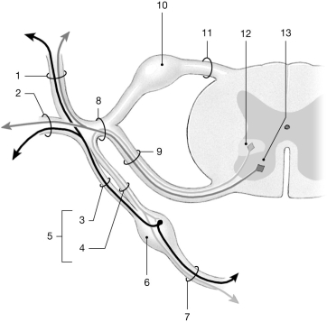
 Figure 13-2 Spinal Nerves
Use Figure 13-2 to answer the following questions:
-Identify the structure labeled "8."
A) peripheral nerve
B) dorsal ramus
C) spinal nerve
D) ventral root
E) dorsal root
G) B) and D)
Correct Answer

verified
Correct Answer
verified
Multiple Choice
 Figure 13-2 Spinal Nerves
Use Figure 13-2 to answer the following questions:
-Identify the structure labeled "4."
A) spinal nerve
B) gray ramus
C) white ramus
D) dorsal ramus
E) ventral ramus
G) D) and E)
Correct Answer

verified
Correct Answer
verified
Multiple Choice
Spinal interneurons inhibit antagonist motor neurons in a process called
A) a crossed extensor reflex.
B) a stretch reflex.
C) a tendon reflex.
D) reciprocal inhibition.
E) reverberating circuits.
G) B) and D)
Correct Answer

verified
Correct Answer
verified
Multiple Choice
The dorsal root of a spinal nerve contains
A) axons of motor neurons.
B) axons of sensory neurons.
C) cell bodies of motor neurons.
D) cell bodies of sensory neurons.
E) interneurons.
G) B) and D)
Correct Answer

verified
Correct Answer
verified
Multiple Choice
The outermost connective-tissue covering of nerves is the
A) endoneurium.
B) endomysium.
C) perineurium.
D) epineurium.
E) epimysium.
G) B) and D)
Correct Answer

verified
Correct Answer
verified
Multiple Choice
 Figure 13-2 Spinal Nerves
Use Figure 13-2 to answer the following questions:
-What is the function of the structure labeled "13"?
A) sensory receptor for pain
B) visceral sensory input
C) somatic motor control
D) somatic sensory input
E) visceral motor control
G) A) and B)
Correct Answer

verified
Correct Answer
verified
Multiple Choice
The white matter of the spinal cord is mainly
A) unmyelinated axons.
B) neuroglia.
C) Schwann cells.
D) myelinated and unmyelinated axons.
E) nodes of Ranvier.
G) B) and C)
Correct Answer

verified
Correct Answer
verified
Showing 81 - 100 of 111
Related Exams